
Cell Culture
Cell Culturing Methods: A Brief History
Discovery of Cells
The term “cell” was first used by the English physicist Robert Hook (1635–1702) in his published work Micrographia in 1665. He describes observations of microscopic structures of biological samples such as insects, plants, sponges, and even fossils. He looked through an early version of a microscope at a thin slice of cork and saw an arrangement of units that he called "cells," as they reminded him of the small rooms (called cellula) that monks used to occupy.
The Dutch scientist Antonie van Leeuwenhoek (1632–1732) improved the microscope design, which allowed him to look at objects at 270× magnifications, while Hooke's microscopes could only achieve about 50× magnifications. This capability allowed him to observe bacteria and protozoa. He was also the first to observe and describe spermatozoa in 1677.
Further improvements to the microscope design helped the botanist Matthias Schleiden (1804–1881) and the zoologist Theodor Schwann (1810–1882) to formulate the "cell theory", which postulates that every organism, whether plant structures or animal tissues, consists of cells. In 1838, Schleiden reasoned that all plant structural elements are made of cells and observed that an organism's growth is due to an increase in cell size and number. In 1839, Schwann concluded from his examinations of animal tissues that "the elementary parts of all tissues are formed from cells." Schwann described that a cell has three essential elements: a nucleus, a fluid content, and a wall (membrane).
However, Schwann and Schleiden misunderstood how cells grow. In 1855, Rudolf Virchow proposed that new cells develop via the scission of existing cells, and he created the aphorism “omnis cellula e cellula," meaning "All cells come from cells." He described the cell as the fundamental unit of life and created the concept that changes in the function of normal cells cause all diseases.
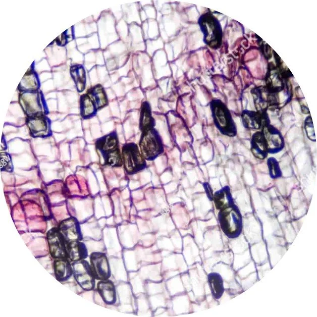
Microscope stained slide of cork cells
Tissue and Cell Culturing
In 1885, the German zoologist Wilhelm Roux (1850 - 1924) established the first tissue culture. He did this by demonstrating that it is possible to maintain living cells (a section of the medullary plate of an embryonic chicken) outside the body in a saline buffer for thirteen days.
In the early 20th century, the American embryologist Ross Granville Harrison (1870–1959) developed the first techniques of cell culture in vitro by growing pieces of frog embryonic tissue outside the body. He placed the tissue in a solution of culture medium on a coverslip, flipped it upside down, and positioned it above a glass slide with a depression in the center. This so-called hanging drop technique---adapted from the microbiological method invented by Robert Koch for bacteria studies---was successfully applied to cell cultures. He subsequently maintained nerve cells in culture and monitored nerve fiber development.

The Hanging Drop Technique
A significant limitation of Harrison's experiments was the typically short time before bacterial contaminations infected the hanging drop. He therefore introduced aseptic techniques in working with cell culture media. Glassware was heated in a flame prior to use. Surgical equipment (e.g., needles, scissors, and forceps) was boiled. Cloths and filter papers were autoclaved. These sterile preparation techniques killed most bacteria and fungi and allowed him to maintain cells in vitro for over five weeks.
Montrose Burrows (1884–1947) and Alexis Carrel (1873–1944) adapted the hanging drop cell culture method using a chicken plasma clot, which was much easier to obtain. It was also more homogenous in quality, making the preparation of in vitro cultures more reliable. They established cell cultures of embryonic and adult tissues (normal adult mammalian and cancerous tissues) of many species that could be maintained in vitro for several months.
They subsequently introduced the idea of continuous culture, i.e., establishing new cultures from the old ones without requiring primary cultures from new tissue explants each time. The results were published in the Journal of the American Medical Association in 1910. The term "tissue culture" was also defined for the first time in 1911 as "a plasmatic medium inoculated with small fragments of living tissues."
Carrel and his coworkers produced and maintained a series of alive and dividing chick heart tissue cultures at the Rockefeller Institute in New York City from 1912 to 1946, 2 years after Carrel's death. As the duration of this culture greatly exceeded the average chick life span, the cells were deemed immortal.
However, in 1961, Leonard Hayflick performed experiments demonstrating a finite lifespan for human cells grown in vitro, calling Carrel's immortality hypothesis into question. Several unsuccessful attempts to culture normal chick somatic cells over a few months further exposed a problem in Carrel's hypothesis. To date, what went wrong with Carrel's immortal tissue cultures is still unknown and remains a matter for speculation.
Classification of Cell Cultures
There are numerous ways to classify cell cultures. One classification often used is according to the origins of the cells used in culture. We differentiate between primary cell culture, secondary cell culture, and cell lines.
Primary Cell Culture:
The cells in primary cultures are obtained directly from normal or malignant, adult or embryonic, tissues or organs. The cells are considered primary until the first passage (subculture), i.e., until they are harvested and reseeded. Primary cultures contain different cell types. They are also used extensively in physiology, cytogenetics, pharmacology, tissue engineering, and other fields. The cell lines established from normal tissues display finite growth. On the contrary, cell lines obtained from cancerous tissues could proliferate indefinitely. We can further classify primary cultures as adherent, when cells are grown while attached to a substrate as monolayers, or suspension cell cultures, when cells are free floating in the culture medium.
Secondary Cell Culture:
When primary cells are transferred from one culture vessel to another, we call this a secondary culture. The passage number of secondary cell culture records the number of times the culture has been subcultured, i.e., harvested and reseeded into multiple 'daughter' cell culture flasks. The subculture technique allowed researchers to obtain cell lines by serial subculture cells from primary cell cultures.
Cell Line Culture:
A cell line is a collection of cells that originate from one cell. Cell lines can be finite or continuous. A continuous cell line has acquired the ability to proliferate indefinitely through genetic mutations or artificial modifications. A finite cell line stops dividing (i.e., senesce) after being sub-cultured for 20-80 passages.

Glass microelectrode array (BMSEED)
In-Situ Electrophysiological Readouts in Cell Cultures
Cell culturing techniques vastly improved during the 20th century. However, in situ and non-destructive (i.e., keeping the cells alive) methods beyond microscopy for obtaining functional readouts from cells were challenging.
In 1972, Thomas et al. published the first paper on applying a multielectrode array (MEA) for recording electrical activity from cultured cells. The array consisted of gold electrodes and leads with an adhesion layer on a glass substrate. The leads were insulated with a photoresist. Meanwhile, the electrodes were plated with Platinum black to reduce their impedance and noise during recording sessions. This first MEA consisted of two rows of 15 electrodes. The electrodes were spaced 100 microns apart, and each electrode had a diameter of 7 microns
Many variations of this initial design have been reported in the literature in the following decades, and commercial MEAs were made available by several companies in the 1990s. The fundamental design, however, has changed little.
Biomechanics and Cell Cultures
In traditional cell cultures, the cells are typically grown in a monolayer on glass or plastic substrates. These conditions do not accurately represent the environments that the cells experience in vivo. Importantly, the mechanical stretch of the cells in vivo affects their gene expression and phenotype via mechanotransduction.
Failing to consider the effect of mechanical forces and biomechanics in cell culture experiments in vitro could cause results to differ from the behavior of the studied cells in vivo. Therefore, not considering mechanical forces in cell culture experiments is a cause for the failure of pre-clinical research to predict clinical outcomes, thus contributing to the lack of success in more than 9 out of 10 clinical trials.
We distinguish between physiological and pathological mechanical stretch. Jufri et al. discussed these in their review article.
Cells experience physiological stretch under natural conditions. For example, heart and lung cells expand and compress with each heartbeat and breath, respectively. The strains are typically less than 10%, and the stretch is usually slow (i.e., low strain rate). These mechanical forces are critical to the proper function of the cell. A physiological stretch is often modeled as linear or radial 5-10% strains (higher on some sections in the peripheral nervous system) at a frequency of 0.5-2 Hz over thousands of cycles.
Pathological stretch is a deformation beyond the healthy limit of the cell and causes an injury or trauma. For example, a pathological stretch of cells in the brain initiates the pathophysiological cascade responsible for about 80% of traumatic brain injury (TBI) cases. TBI is also a significant risk factor for neurodegenerative diseases like Alzheimer's Disease. A pathological stretch is modeled as a linear or radial stretch that is often larger than 20% strain, depending on the type of cells, and often occurs only once at high strain rates of >50/s.
The first commercial cell stretching devices for in vitro experimentation were available in the late 1980s and are now offered by several companies.

Stretchable microelectrode array (BMSEED)
In-Situ Electrophysiology and Biomechanics in Cell Cultures: the MEASSuRE System
Before BMSEED developed and commercialized the stretchable microelectrode array (sMEA) technology, electrophysiology and biomechanics were incompatible paradigms. The materials used previously in electrophysiology experiments are notoriously fragile and delicate, and would get damaged or damage the cells upon mechanical deformation. However, ignoring biomechanics in electrophysiology experiments sacrifices physiological relevance because mechanical cues affect cellular gene expression and phenotype.
The invention of sMEAs, the consumables in BMSEED’s MEASSuRE platform, enables researchers to combine electrophysiology with biomechanics. The electrodes on the sMEA stretch with the cells, and enable extracellular recording and stimulation of evoked activity before, during and after stretching. Different models of MEASSuRE are available to mimic physiological and pathological biomechanics in a controlled in-vitro environment.

A cell-stretching apparatus to apply physiological and mechanical stretch
A data-acquisition module for extracellular electrophysiology to assess the cell’s health, function, and maturity before and after stretching
A live-cell imaging system to visualize cells and cellular processes during the injury
These three paradigms are applied concurrently and independently. Currently, BMSEED is the only company offering stretchable microelectrodes for in vitro research applications.
The overall goal of the MEASSuRE system is to make cells in a dish (in vitro) behave more similarly to the same type of cells inside the body (in vivo). Doing so will enhance the accuracy of pre-clinical research and ultimately improve the success rate of clinical trials in developing treatments for complex diseases.
Power Your Research With MEASSuRE
MEASSuRE is the only research tool that enables researchers to independently and simultaneously apply mechanical stretch, electrophysiology, and imaging. Contact BMSEED to learn more about MEASSuRE.
At BMSEED, our mission is to drive innovation and positively impact the biomedical research community.